Impacted Teeth
An impacted tooth is a tooth which has not erupted fully into the mouth and has perhaps got stuck along the way. The most common tooth to become impacted is the wisdom tooth.
Other teeth can become impacted at various times in both adults and children. Some impacted teeth can become associated with decay or localised infection or both. Wisdom teeth which have become affected by more than one episode of infection may well justify surgical removal.
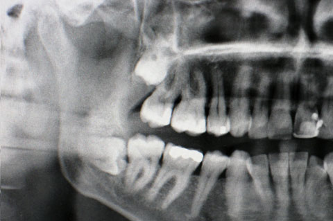
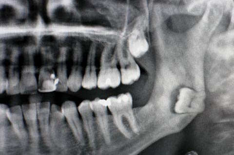
In the case of wisdom teeth surgery, investigation includes taking an X-ray called an OPG (orthopantomograph). This allows the surgeon and patient to see where the wisdom tooth is.
There are two nerves close to an impacted lower wisdom tooth: these are the mandibular nerve, which runs in a tunnel within the mandible (lower jaw) and which supplies feeling to the teeth and also to the lower lip on that side; and the lingual nerve which sits against the inner surface of the lower jaw in the wisdom tooth area before going along the floor of the mouth towards the tongue. It supplies taste and feeling to that side of the tongue. Either or both nerves can be damaged during wisdom tooth surgery.


If an OPG X-ray shows a close relationship of the roots of a wisdom tooth to the mandibular nerve, then further imaging can be important and the best type of imaging is a Cone Beam CT scan (CBCT, a low-volume CT scan in which the patient stands up to have the scan taken).
Surgical removal of impacted wisdom teeth can be done under local anaesthesia, local anaesthesia with sedation or with general anaesthesia. The operation takes approximately 30-45 minutes and may be done on a day case basis. Patients may require up to one week off work, depending on their occupation. Antibiotics are often prescribed for the healing period, together with painkillers and antiseptic mouthwash (Corsodyl).